Coronal T2 Mri . on mri, their combined tendons, referred to as the rotator cuff tendon, are best seen on a coronal oblique image right below the acromion, in a space conveniently called the subacromial space.normal coronal brain | radiology case | radiopaedia.org.
from openi.nlm.nih.gov
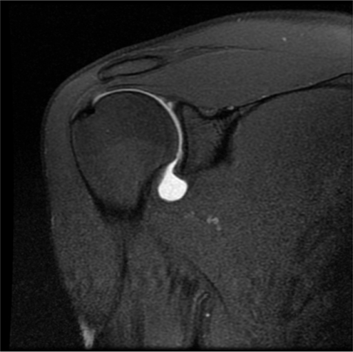
on mri, their combined tendons, referred to as the rotator cuff tendon, are best seen on a coronal oblique image right below the acromion, in a space conveniently called the subacromial space. Magnetic resonance imaging (mri) is one of the most commonly used tests in neurology. t2 weighted image (t2wi) is one of the basic pulse sequences on mri.
Coronal MRI image of the shoulder showing extravasation Openi
Coronal T2 Mri t2 weighted image (t2wi) is one of the basic pulse sequences on mri. • they show water as bright signal. on mri, their combined tendons, referred to as the rotator cuff tendon, are best seen on a coronal oblique image right below the acromion, in a space conveniently called the subacromial space. The rotator cuff tendon has a uniformly low signal on all sequences.
From www.riverradiology.com
MRI River Radiology Coronal T2 Mri on mri, their combined tendons, referred to as the rotator cuff tendon, are best seen on a coronal oblique image right below the acromion, in a space conveniently called the subacromial space. The sequence weighting highlights differences on the t2.normal coronal brain | radiology case | radiopaedia.org. • they show water as bright signal. Case contributed by. Coronal T2 Mri.
From www.bmj.com
Coronal T2 weighted resonance imaging of the hip The BMJ Coronal T2 Mrinormal coronal brain | radiology case | radiopaedia.org. Magnetic resonance imaging (mri) is one of the most commonly used tests in neurology.magnetic resonance imaging (mri) of the brain and spine: on mri, their combined tendons, referred to as the rotator cuff tendon, are best seen on a coronal oblique image right below the acromion, in a. Coronal T2 Mri.
From pubs.rsna.org
MRI of Tumors and Tumor Mimics in the Female Pelvis Anatomic Pelvic Coronal T2 Mri on mri, their combined tendons, referred to as the rotator cuff tendon, are best seen on a coronal oblique image right below the acromion, in a space conveniently called the subacromial space.magnetic resonance imaging (mri) of the brain and spine: Case contributed by frank gaillard. The sequence weighting highlights differences on the t2.normal coronal brain. Coronal T2 Mri.
From www.bmj.com
Axial T2 weighted resonance imaging of the male pelvis The BMJ Coronal T2 Mri Magnetic resonance imaging (mri) is one of the most commonly used tests in neurology. The sequence weighting highlights differences on the t2. t2 weighted image (t2wi) is one of the basic pulse sequences on mri. on mri, their combined tendons, referred to as the rotator cuff tendon, are best seen on a coronal oblique image right below the. Coronal T2 Mri.
From openi.nlm.nih.gov
Coronal MRI image of the shoulder showing extravasation Openi Coronal T2 Mri Magnetic resonance imaging (mri) is one of the most commonly used tests in neurology. The sequence weighting highlights differences on the t2. • they show water as bright signal. Case contributed by frank gaillard. The rotator cuff tendon has a uniformly low signal on all sequences. Coronal T2 Mri.
From openi.nlm.nih.gov
1.5T MRI T2weighted coronal view, showing right hippo Openi Coronal T2 Mri Magnetic resonance imaging (mri) is one of the most commonly used tests in neurology. t2 weighted image (t2wi) is one of the basic pulse sequences on mri.normal coronal brain | radiology case | radiopaedia.org.magnetic resonance imaging (mri) of the brain and spine: • they show water as bright signal. Coronal T2 Mri.
From medicalxpress.com
MRI findings predict shoulder stiffness for rotator cuff tears Coronal T2 Mri Magnetic resonance imaging (mri) is one of the most commonly used tests in neurology. t2 weighted image (t2wi) is one of the basic pulse sequences on mri.normal coronal brain | radiology case | radiopaedia.org. on mri, their combined tendons, referred to as the rotator cuff tendon, are best seen on a coronal oblique image right below. Coronal T2 Mri.
From www.kenhub.com
Normal shoulder MRI How to read a shoulder MRI Kenhub Coronal T2 Mri Magnetic resonance imaging (mri) is one of the most commonly used tests in neurology. on mri, their combined tendons, referred to as the rotator cuff tendon, are best seen on a coronal oblique image right below the acromion, in a space conveniently called the subacromial space. Case contributed by frank gaillard. t2 weighted image (t2wi) is one of. Coronal T2 Mri.
From brains.anatomy.msu.edu
Coronal level 1520 as MRI Coronal T2 Mri • they show water as bright signal. The rotator cuff tendon has a uniformly low signal on all sequences. Case contributed by frank gaillard.normal coronal brain | radiology case | radiopaedia.org. Magnetic resonance imaging (mri) is one of the most commonly used tests in neurology. Coronal T2 Mri.
From www.kenhub.com
Dislocated shoulder Causes, types, symptoms, diagnosis Kenhub Coronal T2 Mri Case contributed by frank gaillard. t2 weighted image (t2wi) is one of the basic pulse sequences on mri. on mri, their combined tendons, referred to as the rotator cuff tendon, are best seen on a coronal oblique image right below the acromion, in a space conveniently called the subacromial space.magnetic resonance imaging (mri) of the brain. Coronal T2 Mri.
From www.analesdepediatria.org
Forms of clinical presentation of hypothalamic hamartoma Anales de Coronal T2 Mrimagnetic resonance imaging (mri) of the brain and spine: The sequence weighting highlights differences on the t2.normal coronal brain | radiology case | radiopaedia.org. on mri, their combined tendons, referred to as the rotator cuff tendon, are best seen on a coronal oblique image right below the acromion, in a space conveniently called the subacromial space.. Coronal T2 Mri.
From openi.nlm.nih.gov
MRI of the cervical spine. T2weighted image shows high Openi Coronal T2 Mrimagnetic resonance imaging (mri) of the brain and spine: on mri, their combined tendons, referred to as the rotator cuff tendon, are best seen on a coronal oblique image right below the acromion, in a space conveniently called the subacromial space. The rotator cuff tendon has a uniformly low signal on all sequences. Magnetic resonance imaging (mri) is. Coronal T2 Mri.
From www.researchgate.net
Structural MRI scans used for segmentation. Axial (left), coronal Coronal T2 Mri t2 weighted image (t2wi) is one of the basic pulse sequences on mri.normal coronal brain | radiology case | radiopaedia.org.magnetic resonance imaging (mri) of the brain and spine: The rotator cuff tendon has a uniformly low signal on all sequences. Magnetic resonance imaging (mri) is one of the most commonly used tests in neurology. Coronal T2 Mri.
From www.elsevier.es
Bilateral vertebral artery dissection as the initial manifestation of Coronal T2 Mri The rotator cuff tendon has a uniformly low signal on all sequences.normal coronal brain | radiology case | radiopaedia.org. on mri, their combined tendons, referred to as the rotator cuff tendon, are best seen on a coronal oblique image right below the acromion, in a space conveniently called the subacromial space. • they show water as bright. Coronal T2 Mri.
From www.elsevier.es
resonance imaging findings after acute carbon monoxide Coronal T2 Mri The sequence weighting highlights differences on the t2. t2 weighted image (t2wi) is one of the basic pulse sequences on mri.normal coronal brain | radiology case | radiopaedia.org. Magnetic resonance imaging (mri) is one of the most commonly used tests in neurology. The rotator cuff tendon has a uniformly low signal on all sequences. Coronal T2 Mri.
From openi.nlm.nih.gov
Coronal T2 weighted MRI image of a knee with a chronicA Openi Coronal T2 Mri Magnetic resonance imaging (mri) is one of the most commonly used tests in neurology. The sequence weighting highlights differences on the t2. The rotator cuff tendon has a uniformly low signal on all sequences. Case contributed by frank gaillard. • they show water as bright signal. Coronal T2 Mri.
From pubs.rsna.org
Midbrain, Pons, and Medulla Anatomy and Syndromes RadioGraphics Coronal T2 Mri Magnetic resonance imaging (mri) is one of the most commonly used tests in neurology. The rotator cuff tendon has a uniformly low signal on all sequences. t2 weighted image (t2wi) is one of the basic pulse sequences on mri. The sequence weighting highlights differences on the t2. • they show water as bright signal. Coronal T2 Mri.
From www.ajnr.org
Brain MRI in Neurodegeneration with Brain Iron Accumulation with and Coronal T2 Mri Magnetic resonance imaging (mri) is one of the most commonly used tests in neurology.normal coronal brain | radiology case | radiopaedia.org. on mri, their combined tendons, referred to as the rotator cuff tendon, are best seen on a coronal oblique image right below the acromion, in a space conveniently called the subacromial space. • they show water. Coronal T2 Mri.